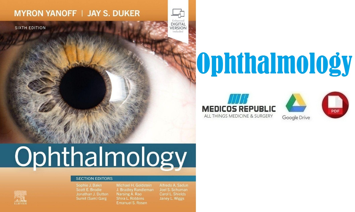
In this article, we are sharing with our audience the genuine PDF download of Ophthalmology 6th Edition PDF using direct links which can be found at the end of this blog post. To ensure user safety and faster downloads, we have uploaded this .pdf file to our online cloud repository so that you can enjoy a hassle-free downloading experience.
Here, at the Medicos Republic, we believe in quality and speed which are a part of our core philosophy and promise to our readers. We hope that you people benefit from our blog! 🙂 Now before we share the free PDF download of Ophthalmology 6th Edition PDF with you, let’s take a look at a few of the important details regarding this ebook.
Overview
Here’s the complete overview of Ophthalmology 6th Edition PDF:
Through five highly regarded editions, Ophthalmology, by Drs. Myron Yanoff and Jay S. Duker, has remained one of the premier texts in the field, providing authoritative guidance on virtually any ophthalmic condition and procedure you may encounter. The fully revised, 6th edition of this award-winning title continues to offer detailed, superbly illustrated content from cover to cover, with extensive updates throughout to keep you current with the latest advancements and fundamentals throughout every subspecialty area in the field. An easy-to-follow, templated format, convenient single volume, and coverage of both common and rare disorders make this title a must-have resource no matter what your level of experience.
Features of Ophthalmology 6th Edition PDF
Here’s a quick overview of the essential features of this book:
- Offers truly comprehensive coverage, including basic foundations through diagnosis and treatment advances across all subspecialties: genetics, optics, refractive surgery, lens and cataract, cornea, retina, uveitis, tumors, glaucoma, neuro-ophthalmology, pediatric and adult strabismus, and oculoplastics.
- Features streamlined, templated chapters, a user-friendly visual layout, and key features boxes for quick access to clinically relevant information and rapid understanding of any topic.
- Contains four new chapters covering Phototherapeutic Keratectomy; IOL Optics; Bag-in-the-lens Cataract Surgery; and Capsulectomy: Modern devices apart from FLACS.
- Includes a fully revised and updated chapter on refractive surgery screening and corneal imaging, as well as an expanded chapter on corneal cross-linking.
- Provides up-to-date information on the latest advances in the field, including new therapies for retinoblastoma, such as intravenous and intraarterial chemotherapy; less common retinal tumor simulators of retinoblastoma; OCT-Angiography; glaucoma stents; new drug delivery platforms; IOL optics; phototherapeutic keratectomy; intraocular pressure monitoring; and more.
- Includes more than 2,000 high-quality illustrations and an expanded video library with more than 60 clips of diagnostic and surgical techniques, including new videos of nystagmus.
- Contains updated management guidelines for central retinal artery occlusions (CRAO).
- Provides fresh perspectives from new section editors Drs. Carol Shields and Sumit (Sam) Garg.
- Enhanced eBook version included with purchase. Your enhanced eBook allows you to access all of the text, figures, and references from the book on a variety of devices.
Table of Contents
Below is the complete table of contents offered inside Ophthalmology 6th Edition PDF:
-
Instructions for online access
-
Cover image
-
Title page
-
Table of Contents
-
Any screen, Any time, Anywhere
-
Copyright
-
User Guide
-
Color Coding
-
Elsevier eBooks+ Version
-
Video Table of Contents
-
Preface
-
Preface to First Edition
-
Contributors
-
In Memory of Emanuel S. Rosen, MD, FRCSE
-
Acknowledgments
-
Dedication
-
Dedication
-
Part 1: Genetics
-
1.1. Fundamentals of Human Genetics
-
Abstract
-
Dna and the Central Dogma of Human Genetics
-
Human Genome
-
Basic Mendelian Principles
-
Mutations
-
Genes and Phenotypes
-
Patterns of Human Inheritance
-
Molecular Mechanisms of Disease
-
Gene Therapy
-
Key References
-
References
-
1.2. Molecular Genetics of Selected Ocular Disorders
-
Abstract
-
Introduction
-
Dominant Corneal Dystrophies
-
Aniridia, Peters’ Anomaly, and Autosomal Dominant Keratitis
-
Rieger’s Syndrome
-
Juvenile Glaucoma
-
Congenital Glaucoma
-
Nonsyndromic Congenital Cataract
-
Retinitis Pigmentosa
-
Stargardt Disease
-
X-Linked Juvenile Retinoschisis
-
Norrie’s Disease
-
Sorsby’s Macular Dystrophy
-
Gyrate Atrophy
-
Color Vision
-
Retinoblastoma
-
Albinism
-
Leber’s Optic Neuropathy
-
Congenital Fibrosis Syndromes and Disorders of Axon Guidance
-
Autosomal Dominant Optic Atrophy
-
Complex Traits
-
Key References
-
References
-
1.3. Genetic Testing and Genetic Counseling
-
Abstract
-
Genetic Testing
-
Genetic Counseling
-
Key References
-
References
-
Part 2: Optics and Refraction
-
2.1. Light
-
Abstract
-
Introduction
-
Geometrical Optics
-
Basic Stigmatic Optics
-
Astigmatic Optics
-
Wave Properties of Light
-
Key References
-
References
-
2.2. Optics of the Human Eye
-
Abstract
-
Introduction
-
Cornea
-
Pupil
-
Lens
-
Accommodation
-
Retina
-
Refractive Errors
-
Key References
-
References
-
2.3. Clinical Refraction
-
Introduction
-
Visual Acuity
-
Spherical Equivalent
-
Detecting Astigmatism
-
Refracting at Near
-
Additional Subjective Techniques
-
Retinoscopy
-
Key References
-
References
-
2.4. Correction of Refractive Errors
-
Abstract
-
Introduction
-
Spectacle Correction
-
Contact Lenses
-
Intraocular Lenses
-
Keratorefractive Surgery
-
Key References
-
References
-
2.5. Ophthalmic Instruments
-
Abstract
-
Introduction
-
Direct Ophthalmoscope
-
Binocular Indirect Ophthalmoscope
-
Fundus Camera
-
Optos Wide-Field Fundus Imaging
-
Optical Coherence Tomography
-
OCT Angiography
-
Slit-Lamp Biomicroscope
-
Slit-Lamp Fundus Lenses
-
Goldmann Applanation Tonometer
-
Specular Microscope
-
Operating Microscope
-
Keratometer and Corneal Topographer
-
Lensmeter
-
Automated Refractor
-
Magnifying Devices
-
Key References
-
References
-
2.6. Wavefront Optics and Aberrations of the Eye
-
Introduction
-
Ray Aberrations
-
The Wavefront Approach To Aberrations
-
Spherical Aberration
-
Coma
-
Distortion
-
Field Curvature
-
Oblique Astigmatism
-
Higher-Order Aberrations
-
Chromatic Aberration
-
Measurement Of Ocular Aberrations
-
An Overall Perspective On Aberration Theory
-
Key References
-
References
-
Part 3: Refractive Surgery
-
3.1. Current Concepts, Classification, and History of Refractive Surgery
-
Abstract
-
Introduction
-
Classification of Refractive Procedures
-
Summary
-
Key References
-
References
-
3.2. Preoperative Evaluation for Refractive Surgery
-
Abstract
-
Introduction
-
Ophthalmic Examination
-
Ancillary Testing
-
Counseling
-
Key References
-
References
-
3.3. Excimer Laser Surface Ablation: Photorefractive Keratectomy (PRK), Laser-Assisted Subepithelial Keratomileusis (LASEK) and Epi-LASIK
-
Abstract
-
Introduction
-
Ablation Profiles
-
Indications
-
Preoperative Evaluation
-
PRK Surgical Technique
-
Results
-
Complications
-
Conclusions
-
Key References
-
References
-
3.4. Laser-Assisted In Situ Keratomileusis (LASIK)
-
Abstract
-
What is Lasik?
-
Indications
-
General Principles
-
Patient Selection
-
Exclusion Criteria
-
Patient Evaluation
-
Surgical Technique
-
Complications
-
Outcomes
-
Future Directions
-
Considerations After Lasik
-
Key References
-
References
-
3.5. Small Incision Lenticule Extraction
-
Abstract
-
Introduction
-
Femtosecond Laser System
-
Treatment Range for Smile
-
Patient Evaluation
-
Surgical Procedure
-
Complications
-
Higher-Order Aberrations
-
Biomechanical Stability
-
Refractive and Visual Outcome
-
Retreatments
-
Conclusion
-
Key References
-
References
-
3.6. Wavefront and Topography-Based Excimer Laser Refractive Surgery
-
Abstract
-
Introduction
-
Lower- and Higher-Order Aberrations
-
Wavefront Optics
-
Measuring Wavefront Aberrations
-
Wavefront-Optimized Treatments
-
Wavefront-Guided Treatments
-
Topography-Guided Treatments
-
Conclusions
-
Key References
-
References
-
3.7. Phakic Intraocular Lenses
-
Abstract
-
Introduction
-
Posterior Chamber
-
IRIS Fixated
-
Indications
-
Advantages Over Keratorefractive Surgery
-
Advantages Over Refractive Lens Exchange
-
Preoperative Evaluation
-
Surgical Technique
-
Postoperative Evaluation
-
Outcomes
-
Potential Complications
-
Conclusion
-
Key References
-
References
-
3.8. Intrastromal Corneal Ring Segments: Synthetic and CAIRS
-
Abstract
-
Introduction
-
Principle of Action
-
Types of ICRS
-
Indications for ICRS
-
Contraindications for ICRS
-
Surgical Techniques
-
Nomograms, Incision Placements, and ICRS Selection
-
Corneal Allogenic Intrastromal Ring Segments
-
Complications
-
ICRS With Corneal Cross-Linking
-
Conclusion
-
Key References
-
References
-
3.9. Surgical Treatment of Presbyopia
-
Abstract
-
Introduction
-
Optical Considerations for Treating Presbyopia
-
Surgical Procedures
-
Key References
-
References
-
3.10. Excimer Laser Phototherapeutic Keratectomy
-
Abstract
-
Introduction
-
Indications and Contraindications
-
Preoperative Evaluation
-
Surgical Technique
-
Postoperative Care
-
Outcomes
-
Complications
-
Conclusions
-
Key References
-
References
-
3.11. Corneal Cross-linking
-
Abstract
-
Corneal Cross-Linking is a Photochemical Reaction
-
Indication for CXL
-
Biological Interactions and Morphological Changes in CXL
-
Wound Healing and Keratocyte Cytotoxicity in CXL
-
Creating Reactive Oxygen Species at Deeper Levels with CXL
-
Biological Differences Associated with Changes in Irradiation Time in CXL
-
Role of the Riboflavin Carrier in CXL
-
The Role of LYSYL Oxidase Enzyme in the Biology of CXL
-
CXL in Thin Corneas
-
Photoactivated Chromophore Treatment for Corneal Cross-Linked Infectious Keratitis
-
Corneal Cross-Linking Combined with Refractive Surgical Procedures
-
Corneal Haze and Infiltrates After CXL
-
Conclusion
-
Key References
-
References
-
Part 4: Cornea and Ocular Surface Diseases
-
Section 1: Basic Principles
-
4.1. Corneal Anatomy, Physiology, and Wound Healing
-
Abstract
-
Introduction
-
Corneal Wound Healing
-
Key References
-
References
-
4.2. Anterior Segment Imaging Modalities
-
Abstract
-
Introduction
-
Anterior Segment Optical Coherence Tomography
-
Specular Microscopy
-
Ultrasound Biomicroscopy
-
Meibography
-
In Vivo Confocal Microscopy
-
Topography And Tomography
-
Wavefront Analysis
-
Summary
-
Key References
-
References
-
Section 2: Congenital Abnormalities
-
4.3. Congenital Corneal Anomalies
-
Abstract
-
Introduction
-
Size And Shape Anomalies
-
Anomalies Of Corneal Clarity
-
Acknowledgment
-
Key References
-
References
-
Section 3: External Diseases
-
4.4. Blepharitis
-
Abstract
-
Introduction
-
Epidemiology
-
Pathogenesis
-
Ocular Manifestations
-
Diagnosis And Ancillary Testing
-
Treatment
-
Key References
-
References
-
4.5. Herpes Zoster Ophthalmicus
-
Abstract
-
Epidemiology And Pathogenesis
-
Clinical Manifestations
-
Herpes Zoster Ophthalmicus In Acquired Immune Deficiency Syndrome
-
Diagnosis
-
Management
-
Prevention
-
Key References
-
References
-
Section 4: Conjunctival Diseases
-
4.6. Infectious Conjunctivitis
-
Abstract
-
Infectious Conjunctivitis
-
Noninfectious Conjunctivitis
-
Key References
-
References
-
4.7. Allergic Conjunctivitis
-
Abstract
-
Acute Allergic Conjunctivitis: Seasonal/Perennial
-
Chronic Atopic Keratoconjunctivitis
-
Vernal Keratoconjunctivitis
-
Treatment Of Allergic/Atopic Keratoconjunctivitis
-
Allergic Dermatoconjunctivitis
-
Microbial Allergic Conjunctivitis
-
Giant Papillary Conjunctivitis
-
Dupilumab-Associated Conjunctivitis
-
Key References
-
References
-
4.8. Tumors of the Conjunctiva
-
Abstract
-
Tumors of the Conjunctiva
-
Ocular Surface Squamous Neoplasia
-
Key References
-
References
-
4.9. Pterygium and Conjunctival Degenerations
-
Abstract
-
Introduction
-
Pinguecula
-
Pterygium
-
Senile Scleral Plaques
-
Conjunctival Amyloid
-
Conjunctival Melanosis
-
Key References
-
References
-
4.10. Ocular Cicatricial Pemphigoid/Mucous Membrane Pemphigoid
-
Abstract
-
Introduction
-
Pathogenesis
-
Clinical Findings
-
Diagnosis
-
Treatment
-
Conclusion
-
Key References
-
References
-
Section 5: Scleral and Episcleral Diseases
-
4.11. Episcleritis and Scleritis
-
Abstract
-
Introduction
-
Inflammatory Diseases
-
Key References
-
References
-
Section 6: Corneal Diseases
Ophthalmology 6th Edition PDF Free Download
Alright, now in this part of the article, you will be able to access the free PDF download of Ophthalmology 6th Edition PDF using our direct links mentioned at the end of this article. We have uploaded a genuine PDF ebook copy of this book to our online file repository so that you can enjoy a blazing-fast and safe downloading experience.
[adinserter block=”3″]
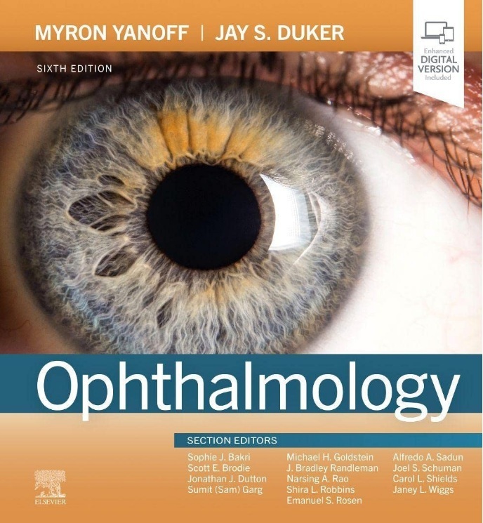
Here’s the cover image preview of Ophthalmology 6th Edition PDF:

FILE SIZE: 44 MB
[adinserter block=”2″]
Please use the direct link mentioned below to download Ophthalmology 6th Edition PDF for free now:
Download Link
Happy learning, people! 🙂

DMCA Disclaimer: This site complies with DMCA Digital Copyright Laws.
PLEASE NOTE: We do not host/store any copyrighted content on our website, it’s a catalog of links that are already found on the internet. Please check out our DMCA Policy. If you feel that we have violated your copyrights, please get in touch with us immediately, and the said content will be PERMANENTLY removed within 24 hours.
You may send an email to madxperts [at] gmail.com for all DMCA / Removal Requests or use our Contact Us page.
Check out our DMCA Policy.


![The LASIK Handbook: A Case-Based Approach PDF Free Download [Direct Link] The LASIK Handbook A Case-Based Approach PDF](https://www.medicosrepublic.com/wp-content/uploads/2024/02/The-LASIK-Handbook-A-Case-Based-Approach-PDF-218x150.jpg)
![Refractive Surgery 3rd Edition PDF Free Download [Direct Link] Refractive Surgery 3rd Edition PDF](https://www.medicosrepublic.com/wp-content/uploads/2024/02/Refractive-Surgery-3rd-Edition-PDF-218x150.jpg)
![The Little Eye Book: A Pupil’s Guide to Understanding Ophthalmology PDF Free Download [Direct Link]](https://www.medicosrepublic.com/wp-content/uploads/2024/02/The-Little-Eye-Book-A-Pupils-Guide-to-Understanding-Ophthalmology-PDF-218x150.jpg)
![OphthoBook Questions PDF Free Download [Direct Link] OphthoBook Questions PDF](https://www.medicosrepublic.com/wp-content/uploads/2024/02/OphthoBook-Questions-PDF-218x150.jpg)
![OphthoBook PDF Free Download [Direct Link]](https://www.medicosrepublic.com/wp-content/uploads/2024/02/OphthoBook-PDF-218x150.jpg)
![Eye Anatomy Coloring Book PDF Free Download [Direct Link]](https://www.medicosrepublic.com/wp-content/uploads/2024/02/Eye-Anatomy-Coloring-Book-PDF-218x150.jpg)
![Kaplan Medical USMLE Pharmacology and Treatment Flashcards PDF Free Download [Direct Link] Kaplan Medical USMLE Pharmacology and Treatment Flashcards PDF](https://www.medicosrepublic.com/wp-content/uploads/2023/01/Kaplan-Medical-USMLE-Pharmacology-and-Treatment-Flashcards-PDF-Free-Download-150x150.jpg)
![NCLEX-RN Prep Plus 2018: 2 Practice Tests + Proven Strategies + Online + Video PDF Free Download [Direct Link] Kaplan's NCLEX-RN Prep Plus 2018 PDF](https://www.medicosrepublic.com/wp-content/uploads/2018/08/Kaplans-NCLEX-RN-Prep-Plus-2018-PDF-Free-Download-150x150.jpg)
![Pocket Medicine 5th Edition PDF Free Download [Direct Link] Pocket Medicine 5th Edition PDF](https://www.medicosrepublic.com/wp-content/uploads/2022/10/Pocket-Medicine-5th-Edition-PDF-Free-Download-150x150.jpg)
![DK Encyclopedia of Herbal Medicine PDF Free Download [Direct Link] DK Encyclopedia of Herbal Medicine PDF](https://www.medicosrepublic.com/wp-content/uploads/2024/03/DK-Encyclopedia-of-Herbal-Medicine-PDF-Free-Download-1-150x150.jpg)
![Leadership and the Advanced Practice Nurse: The Future of a Changing Healthcare Environment PDF Free Download [Direct Link] Leadership and the Advanced Practice Nurse: The Future of a Changing Healthcare Environment PDF](https://www.medicosrepublic.com/wp-content/uploads/2023/04/Leadership-and-the-Advanced-Practice-Nurse-The-Future-of-a-Changing-Healthcare-Environment-PDF-Free-150x150.jpg)
![Master the Wards: Internal Medicine Flashcards PDF Free Download [Direct Link]](https://www.medicosrepublic.com/wp-content/uploads/2022/06/Master-the-Wards-Internal-Medicine-Flashcards-PDF-Free-Download-696x368-1-150x150.jpg)
![The Green Pharmacy PDF Free Download [Direct Link] The Green Pharmacy PDF](https://www.medicosrepublic.com/wp-content/uploads/2024/03/The-Green-Pharmacy-PDF-Free-Download-1-150x150.jpg)
![Oral Microbiology and Immunology 3rd Edition PDF Free Download [Direct Link] Oral Microbiology and Immunology 3rd Edition PDF](https://www.medicosrepublic.com/wp-content/uploads/2023/03/Oral-Microbiology-and-Immunology-3rd-Edition-PDF-Free-150x150.jpg)
![Dermatology an illustrated color text PDF Free Download [Direct Link] Dermatology an illustrated color text PDF](https://www.medicosrepublic.com/wp-content/uploads/2022/05/Dermatology-an-illustrated-color-text-PDF-Free-Download-150x150.jpg)
![General Anatomy and Musculoskeletal System (THIEME Atlas of Anatomy) 1st Edition PDF Free Download [Direct Link] General Anatomy and Musculoskeletal System (THIEME Atlas of Anatomy) 1st Edition PDF](https://www.medicosrepublic.com/wp-content/uploads/2022/09/General-Anatomy-and-Musculoskeletal-System-1st-Edition-PDF-Free-Download-150x150.jpg)


![Carcinoid Syndrome Mnemonic [Easy-to-memorize] Carcinoid Syndrome Mnemonic](https://www.medicosrepublic.com/wp-content/uploads/2024/08/Carcinoid-Syndrome-Mnemonic-Easy-to-memorize-100x70.jpg)

![Color Atlas of Veterinary Histology 3rd Edition PDF Free Download [Direct Link] Color Atlas of Veterinary Histology 3rd Edition PDF](https://www.medicosrepublic.com/wp-content/uploads/2024/07/Color-Atlas-of-Veterinary-Histology-3rd-Edition-PDF-Free-Download-100x70.jpg)