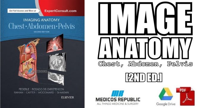
In this article, we are sharing with our audience the genuine PDF download of Imaging Anatomy: Chest, Abdomen, Pelvis 2nd Edition PDF using direct links which can be found at the end of this blog post. To ensure user-safety and faster downloads, we have uploaded this .pdf file to our online cloud repository so that you can enjoy a hassle-free downloading experience.
At Medicos Republic, we believe in quality and speed which are a part of our core philosophy and promise to our readers. We hope that you people benefit from our blog! 🙂
Now before that we share the free PDF download of Imaging Anatomy: Chest, Abdomen, Pelvis 2nd Edition PDF with you, let’s take a look into few of the important details regarding this ebook.
Overview
Here’s the complete overview of Imaging Anatomy: Chest, Abdomen, Pelvis 2nd Edition PDF:
Designed to help you quickly learn or review normal anatomy and confirm variants, Imaging Anatomy: Chest, Abdomen, Pelvis provides detailed views of anatomic structures in successive imaging slices in each standard plane of imaging. Axial, coronal, sagittal, and 3D reconstructions accompany highly accurate and detailed medical drawings, assisting you in making an accurate diagnosis. Comprehensive coverage of the chest, abdomen, and pelvis, combined with an orderly, easy-to-follow structure, make this unique title unmatched in its field.
Features of Imaging Anatomy: Chest, Abdomen, Pelvis 2nd Edition PDF
Here’s a quick overview of the important features of this book:
- Includes all relevant imaging modalities, 3D reconstructions, and highly accurate and detailed medical drawings that illustrate the fine points of the imaging anatomy
- Depicts common anatomic variants and covers common pathological processes as a part of its comprehensive coverage
- Provides a detailed overview of airway and interstitial network anatomy-the basis for understanding and diagnosing interstitial lung disease
- Features representative pathologic examples to highlight the effect of disease on human anatomy
- Includes plain radiography, the latest generation of multi-planar advanced cross-sectional MR and CT, ultrasound for pelvis/renal/liver/gallbladder, barium for GI tract, and much more
- Offers state of the art, detailed pelvic floor imaging and perianal/perirectal fistula imaging using high-resolution CT and MR, including 3T MR
- Expert Consult eBook version included with purchase. This enhanced eBook experience allows you to search all of the text, figures, images, and references from the book on a variety of devices.
Michael P Federle (Author)
By Michael P Federle, MD, FACR, Professor and Associate Chair for Education , Department of Radiology, Stanford University School of Medicine, Stanford, California; Melissa L. Rosado-de-Christenson, MD, FACR, Section Chief, Thoracic Radiology, Department of Radiology, Saint Luke’s Hospital of Kansas City, Professor of Radiology, University of Missouri-Kansas City School of Medicine, Kansas City, Missouri; Akram M. Shaaban, MBBCh, Professor of Radiology, Department of Radiology and Imaging Sciences, University of Utah School of Medicine, Salt Lake City, Utah and Paula J. Woodward, MD, Professor of Radiology, David G. Bragg, MD and Marcia R. Bragg Presidential Endowed Chair in Oncologic Imaging, Adjunct Professor of Obstetrics and Gynecology, University of Utah School of Medicine, Salt Lake City, Utah.
Table of Contents
Below is the complete table of contents offered inside Imaging Anatomy: Chest, Abdomen, Pelvis 2nd Edition PDF:
- Chest Overview
- Lung Development
- Airway Structure
- Vascular Structure
- Interstitial Network
- Lungs
- Hila
- Airways
- Pulmonary Vessels
- Pleura
- Mediastinum
- Systemic Vessels
- Heart
- Coronary Arteries and Cardiac Veins
- Pericardium
- Chest Wall
- ABDOMEN
- Embryology of Abdomen
- Abdominal Wall
- Diaphragm
- Peritoneal Cavity
- Vessels, Lymphatic System and Nerves, Abdominal
- Esophagus
- Gastroduodenal
- Small Intestine
- Colon
- Spleen
- Liver
- Biliary System
- Pancreas
- Retroperitoneum
- Adrenal
- Kidney
- Ureter and Bladder
- PELVIS
- Vessels, Lymphatic System and Nerves, Pelvic
- Male
- Male Pelvic Wall and Floor
- Testes and Scrotum
- Prostate and Seminal Vesicles
- Penis and Urethra
- Female
- Female Pelvic Floor
- Uterus
- Ovaries
You might also be interested in: 🙂
USMLE Step 1 Lecture Notes 2018: Anatomy PDF Free Download
Product Details
Below are the technical specifications of Imaging Anatomy: Chest, Abdomen, Pelvis 2nd Edition PDF:
- Hardcover: 1192 pages
- Publisher: Elsevier; 2 edition (13 Feb. 2017)
- Language: English
- ISBN-10: 032347781X
- ISBN-13: 978-0323477819
- Product Dimensions: 22.2 x 28.1 cm
Imaging Anatomy: Chest, Abdomen, Pelvis 2nd Edition PDF Free Download
Alright, now in this part of the article, you will be able to access the free PDF download of Imaging Anatomy: Chest, Abdomen, Pelvis 2nd Edition PDF using our direct links mentioned at the end of this article. We have uploaded a genuine PDF ebook copy of this book to our online file repository so that you can enjoy a blazing-fast and safe downloading experience.
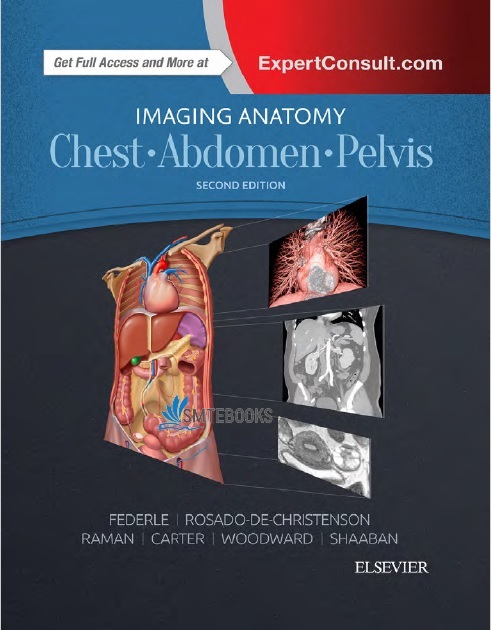
Here’s the cover image preview of Imaging Anatomy: Chest, Abdomen, Pelvis 2nd Edition PDF:
FILE SIZE: 113 MB
Please use the direct link mentioned below to download Imaging Anatomy: Chest, Abdomen, Pelvis 2nd Edition PDF for free now:
Download Link
Happy learning, people!
DMCA Disclaimer: This site complies with DMCA Digital Copyright Laws. Please bear in mind that we do not own copyrights to these books. We’re sharing this material with our audience ONLY for educational purpose. We highly encourage our visitors to purchase original books from the respected publishers. If someone with copyrights wants us to remove this content, please contact us immediately.
All books/videos on the Medicos Republic are free and NOT HOSTED ON OUR WEBSITE. If you feel that we have violated your copyrights, then please contact us immediately (click here).
Check out our DMCA Policy.
You may send an email to madxperts [at] gmail.com for all DMCA / Removal Requests.


![Super Simple Anatomy and Physiology: The Ultimate Learning Tool 2nd Edition PDF Free Download [Direct Link] Super Simple Anatomy and Physiology PDF](https://www.medicosrepublic.com/wp-content/uploads/2023/07/Super-Simple-Anatomy-and-Physiology-PDF-218x150.jpg)
![The Anatomy Coloring Book 2nd Edition PDF Free Download [Direct Link] The Anatomy Coloring Book 2nd Edition PDF](https://www.medicosrepublic.com/wp-content/uploads/2023/07/The-Anatomy-Coloring-Book-2nd-Edition-PDF-218x150.jpg)
![The Body Atlas PDF Free Download [Direct Link] The Body Atlas PDF](https://www.medicosrepublic.com/wp-content/uploads/2023/07/The-Body-Atlas-PDF-218x150.jpg)
![Understanding Anatomy and Physiology 2nd Edition PDF Free Download [Direct Link] Understanding Anatomy and Physiology 2nd Edition PDF](https://www.medicosrepublic.com/wp-content/uploads/2023/07/Understanding-Anatomy-and-Physiology-2nd-Edition-PDF-218x150.jpg)
![Gray. Anatomía para estudiantes PDF Free Download [Direct Link]](https://www.medicosrepublic.com/wp-content/uploads/2023/07/Gray.-Anatomia-para-estudiantes-PDF-218x150.jpg)
![Anatomy and Physiology with Integrated Study Guide 6th Edition PDF Free Download [Direct Link] Anatomy and Physiology with Integrated Study Guide 6th Edition PDF](https://www.medicosrepublic.com/wp-content/uploads/2023/07/Anatomy-and-Physiology-with-Integrated-Study-Guide-6th-Edition-PDF-218x150.jpg)
![Vishram Singh Neuroanatomy PDF Free Download [Direct Link]](https://www.medicosrepublic.com/wp-content/uploads/2022/05/Vishram-Singh-Neuroanatomy-PDF-Free-Download-1-150x150.jpg)
![Cardiovascular Pathology The Perfect Preparation for USMLE Step 1 PDF Free Download [Direct Link]](https://www.medicosrepublic.com/wp-content/uploads/2022/06/Cardiovascular-Pathology-The-Perfect-Preparation-for-USMLE-Step-1-PDF-Free-Download-1-696x365-1-150x150.jpg)
![Rapid Interpretation of EKG’s PDF Free Download [Direct Link] Rapid Interpretation of EKG’s PDF](https://www.medicosrepublic.com/wp-content/uploads/2022/05/Rapid-Interpretation-of-EKG’s-PDF-Free-Download--150x150.jpg)
![Pocketbook of Clinical IR: A Concise Guide to Interventional Radiology PDF Free Download [Direct Link]](https://www.medicosrepublic.com/wp-content/uploads/2023/02/Pocketbook-of-Clinical-IR-A-Concise-Guide-to-Interventional-Radiology-PDF-Free-Download-150x150.jpg)
![The Night Land PDF Free Download [Direct Link] The Night Land PDF Free Download](https://www.medicosrepublic.com/wp-content/uploads/2023/04/The-Night-Land-PDF-Free-Download-150x150.jpg)
![Emergency Responder: Advanced First Aid for Non-EMS Personnel PDF Free Download [Direct Link] Emergency Responder Advanced First Aid for Non-EMS Personnel PDF](https://www.medicosrepublic.com/wp-content/uploads/2023/03/Emergency-Responder-Advanced-First-Aid-for-Non-EMS-Personnel-PDF-150x150.jpg)
![Hutchisons Clinical Methods 24th Edition PDF Free Download [Direct Link]](https://www.medicosrepublic.com/wp-content/uploads/2022/06/Hutchisons-Clinical-Methods-24th-Edition-PDF-Free-Download-696x348-1-150x150.jpg)
![Networking For Dummies PDF Free Download [Direct Link] Networking For Dummies PDF](https://www.medicosrepublic.com/wp-content/uploads/2023/02/Networking-For-Dummies-PDF-150x150.jpg)
![ABC of Emergency Differential Diagnosis 1st Edition PDF Free Download [Direct Link] ABC of Emergency Differential Diagnosis PDF](https://www.medicosrepublic.com/wp-content/uploads/2023/07/ABC-of-Emergency-Differential-Diagnosis-PDF-150x150.jpg)
![Atrial Fibrillation Causes Mnemonic [PIRATES]](https://www.medicosrepublic.com/wp-content/uploads/2023/08/Atrial-Fibrillation-Causes-Mnemonic-PIRATES-150x150.jpg)


